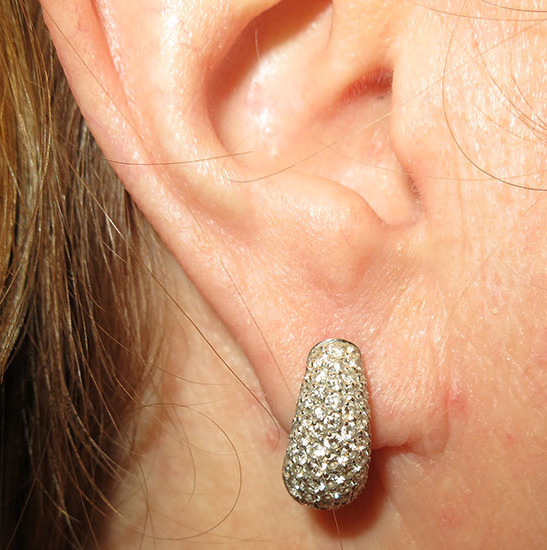
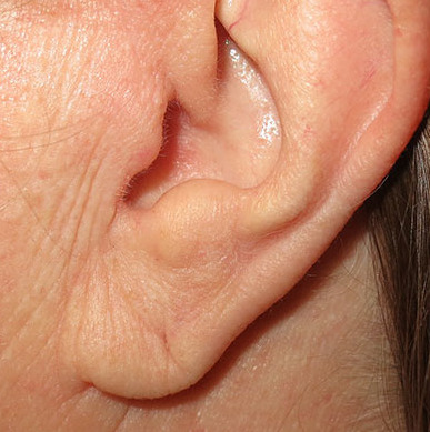
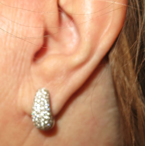
Sign-up for our New Digital Chart System Here

Before and after keloid excision and ear reconstruction in a 24 year old woman. She had previously removed an infected ear piercing and gradually developed a keloid scar on her right ear. It extended from the front all the way through the piercing to the back of her ear.

She had failed conservative treatment including scar therapy and steroid injection. A keloid scar is in fact a “tumor”, which is a mass of tissue that keeps growing beyond its original borders. Keloid scars are benign, but can be painful, itchy and have obvious cosmetic implications. This patient’s keloid scar was excised and her ear reconstructed in the office under local anesthetic. The actual keloid from the front and back are shown as the last images.

Follow up photos are shown 6 weeks after surgery. Scar therapy including use of a topical scar gel and scar massage has just started. This patient’s ear and wound healing progression to mature scar formation will be watched closely going forward, as keloid scars can recur. If she begins to produce too much immature scar tissue (known as a “hypertrophic scar”), then steroid injection can help to soften the scar and switch the body’s scar formation toward mature scar.

As a last resort for recurrent or refractory keloids, a combination of surgical excision and three short bursts of radiation therapy to the scar are usually successful. We hope that this patient continues to heal well without need for any further treatments!

before
after
before
after
before
after

*All photos are actual patient photographs and are for illustrative purposes only. Individual results may vary.

Dr Karen Horton