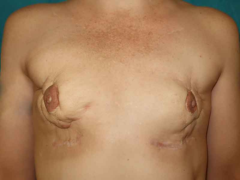
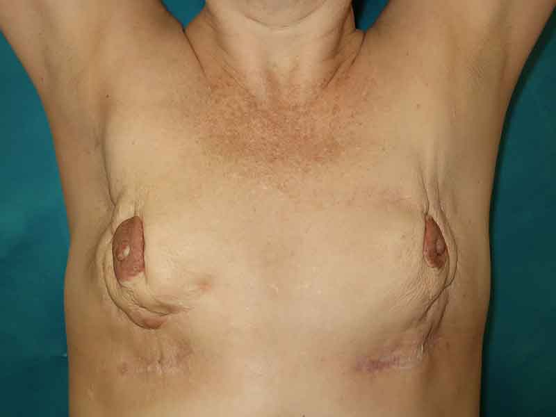
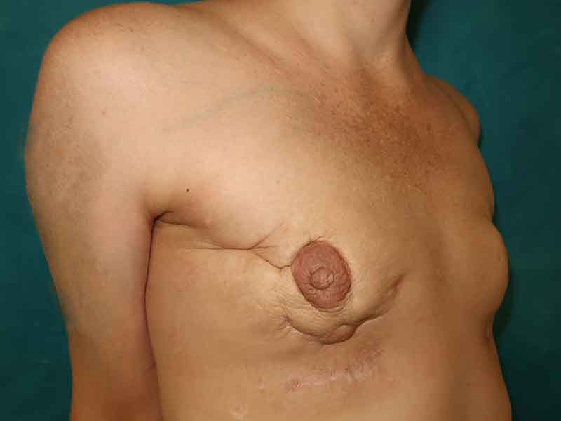
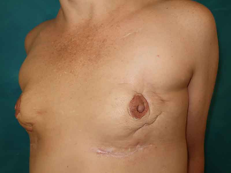
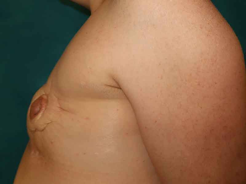
Before and after bilateral delayed breast implant reconstruction in a 48 year old breast cancer survivor. Her past surgical history was extensive; including bilateral mastectomies and tissue expander-submuscular implant reconstruction, chemotherapy, radiation therapy, a breast revision to implants on top of the muscle, chronic infections and the decision to remove both of her implants to give her body a break.
Her implants had been out for well over a year and she had developed chronic seromas that had not been completely absorbed, which filled the pocket and contributed to her preoperative deformity. Although she thought she would be satisfied having no reconstruction at all, she decided she wanted to give breast reconstruction a final attempt.
She had been counselled extensively about her risks of implant related complications should she give an implant based breast reconstruction another try. She was a candidate for a flap, and despite that being our recommendation given her past history, she wanted to give implants one last try.
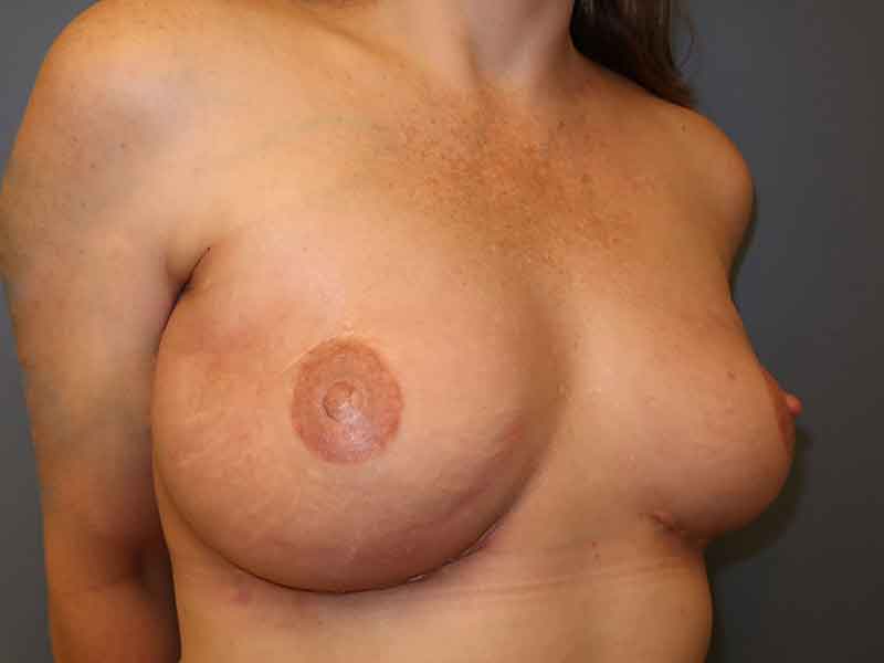
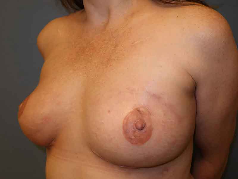
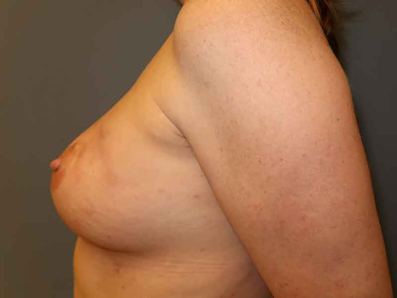
An extensive total capsulectomy removed all the interior scar tissue and drained the old seroma. Cultures were taken of the capsule, as is standard practice with capsular contracture or past implant infection, and these were thankfully negative.
The full sized new smooth round silicone gel breast implants were placed in the space on top of the pectoralis major muscles. No tissue expansion was required, and she was grateful that the implants were not placed under her muscles again.
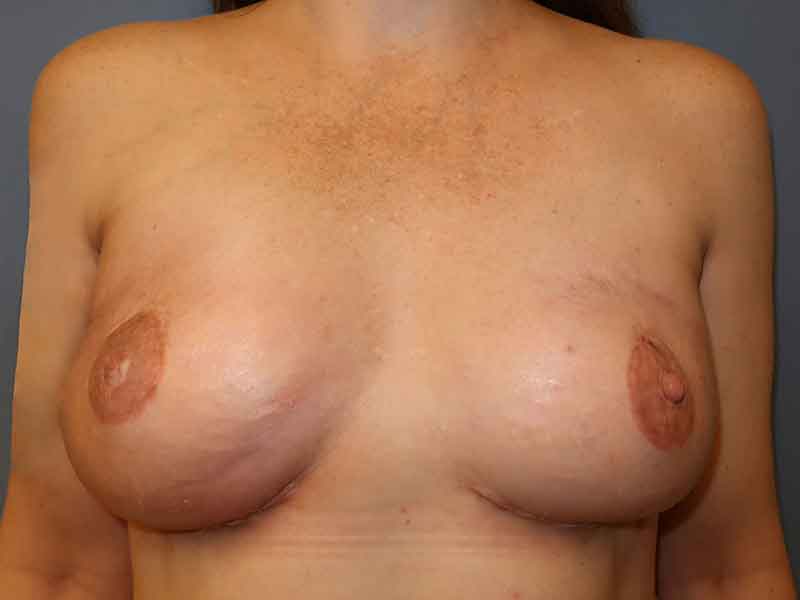
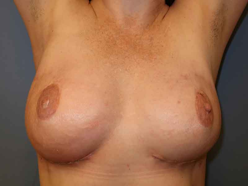
Follow up photos are shown three weeks after surgery. Her incisions are still considered “wounds”, as evidenced by scabbing and stitched-looking incisions. She has residual firm edema of the breast skin that will take several more months to go away.
Her “pre-pectoral” implants (on top of the muscle) have a natural teardrop shape. Her surgery only required 24 hours of pain medication – much different from the lengthy process of tissue expansion under the muscle she went through several years ago. She is very happy she went forward and we are all crossing our fingers that she has no more complications!
*All photos are actual patient photographs and are for illustrative purposes only. Individual results may vary.