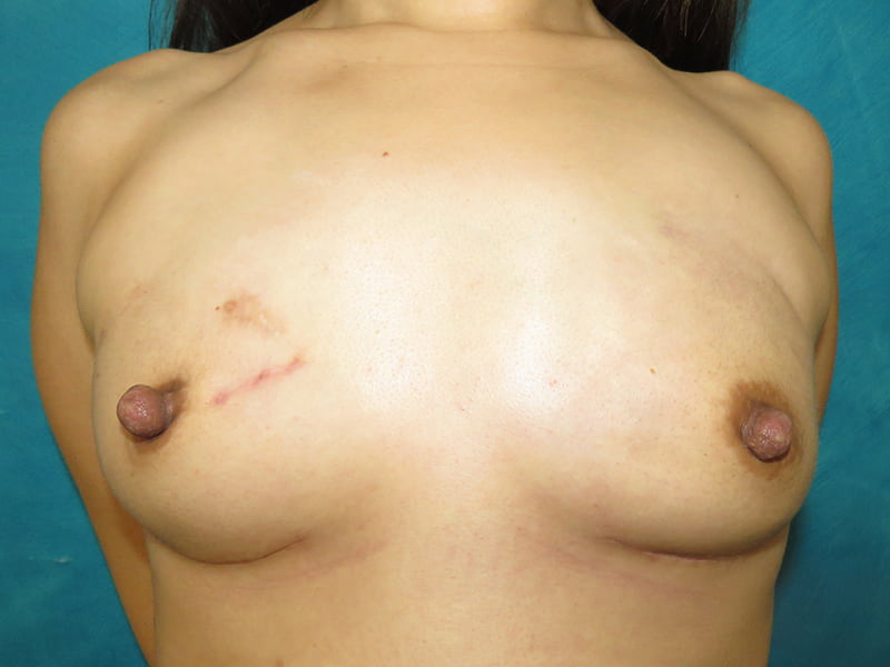
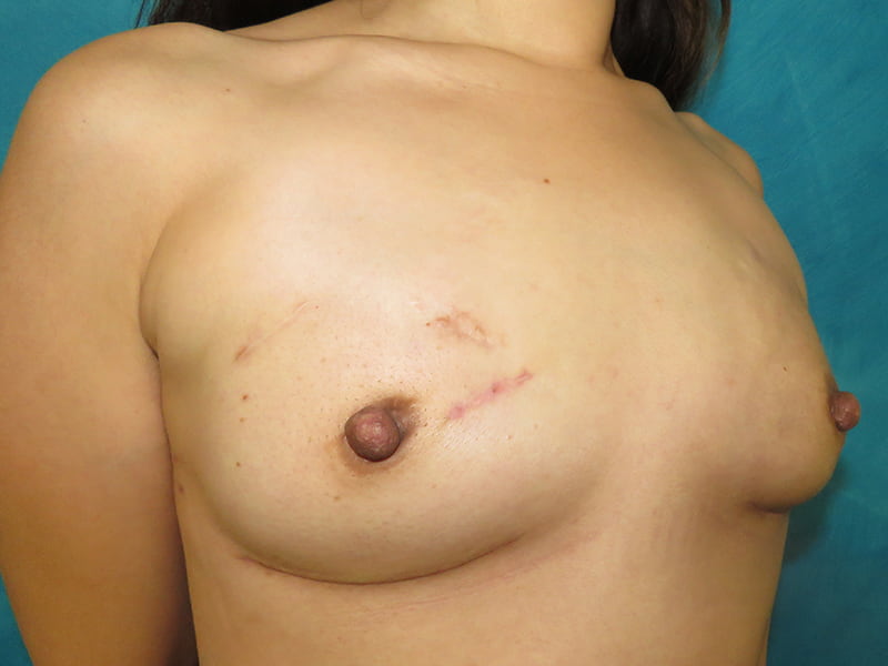
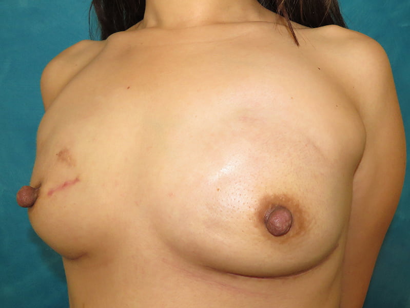
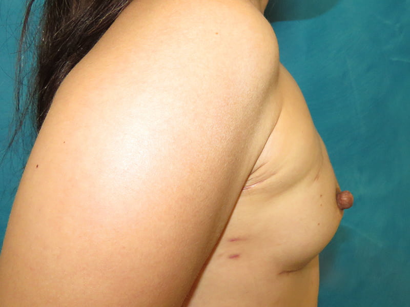
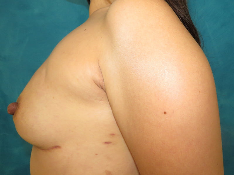
Before and after bilateral breast implant reconstruction revision in a 33-year-old breast cancer survivor. She had previously had three lumpectomies on her right breast and required a completion mastectomy. She chose to have a contralateral prophylactic mastectomy as well on the other breast.
Tissue expanders had been placed, and she was ready for her final implant. She sought our office as she liked the natural look she saw on our before and after galleries and she wanted to be “treated like a patient, not a number.”
She had not yet had children, but felt she deserved a makeover for surviving cancer! She disliked the flat, hard expanders and wanted this surgery to function as her “breast cancer makeover.” Her goals were for her breasts to be larger than her preoperative breast size.
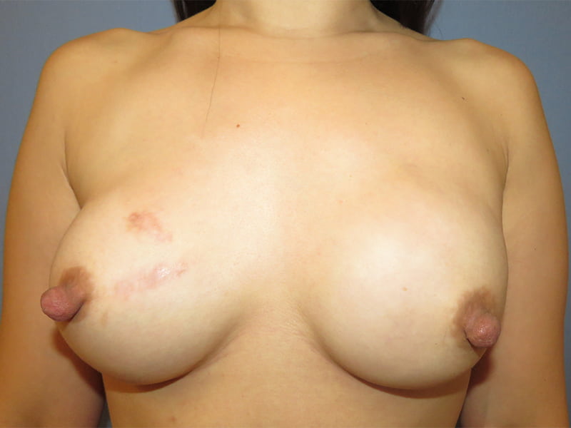
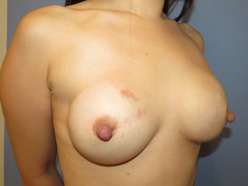
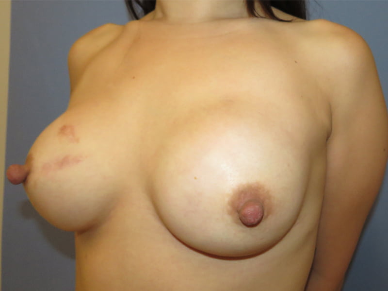
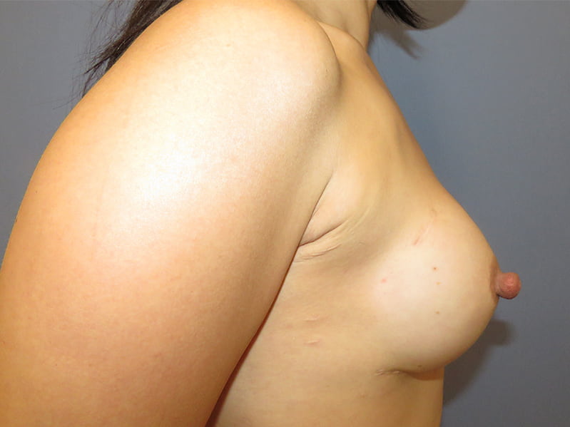
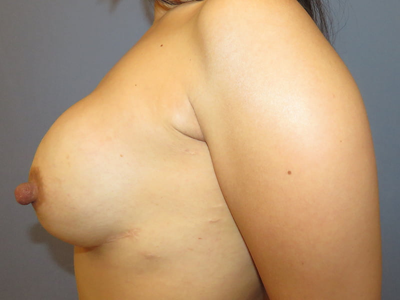
Her tissue expanders were removed, and the pockets were opened to accommodate a fuller, rounder implant. New smooth round silicone gel breast implants were placed on top of the muscle, in the prepectoral position. We always add liposuction to the axillary rolls to any breast procedure when excess fat is present in the armpits and bra rolls.
Long-term follow up photos are shown 2 years after surgery with mature scars and her final results. She will now follow up with us at 5 years postop, 10 years and then every 10 years going forward.
*All photos are actual patient photographs and are for illustrative purposes only. Individual results may vary.