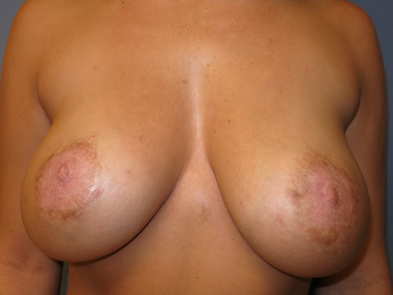
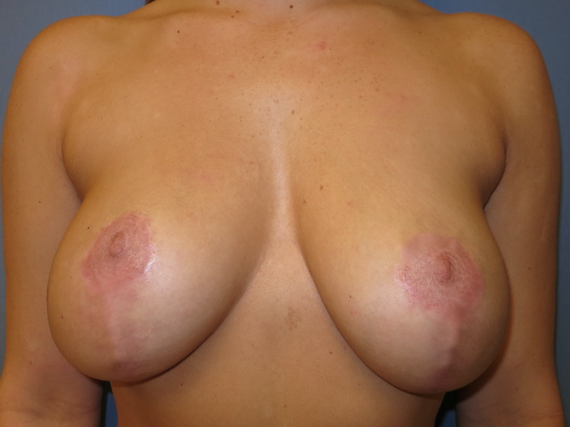
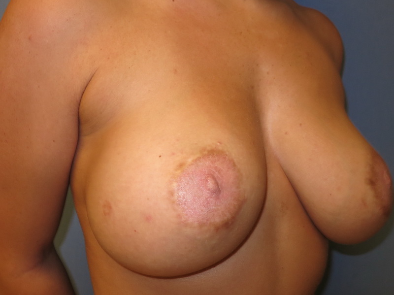
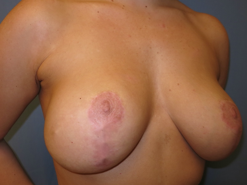
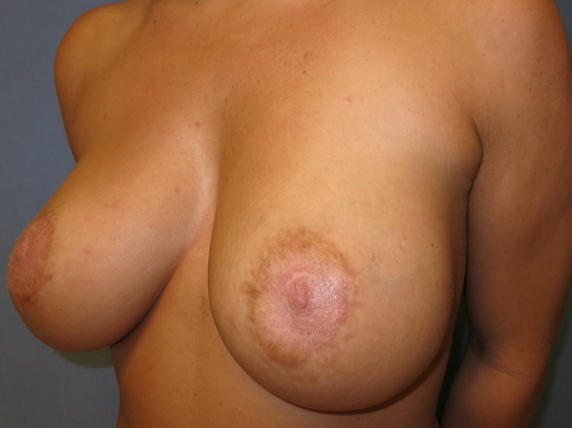
Before and after bilateral breast revision and skin-only mastopexy in a 26 year old woman. She previously had an attempt at an areola reduction and lift by the circumareolar technique, also known as “donut mastopexy” or the Benelli technique. Her scars had hyperpigmented, healing darker than her surrounding skin. This was probably from “post-inflammatory hyperpigmentation”, where tension on the wound caused deposition of excess melanin, the skin pigment.
The problem with trying to reduce the areola diameter and achieving a lift only with the circular incision is related to physics. If there is only a circular incision, there is no relief of tension off the circle (think of a spinning ride at the fair where you get pulled firmly against the wall from centripetal force).
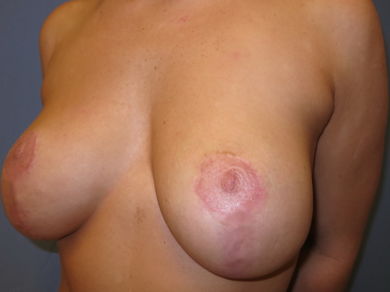
In order to relieve tension on the circle during healing, another incision is required under the breast. In other words, the donut needs to be converted to a lollipop. The vertical incision takes pressure off the circle during healing and prevents areolar spreading. Read this blog post for more information.
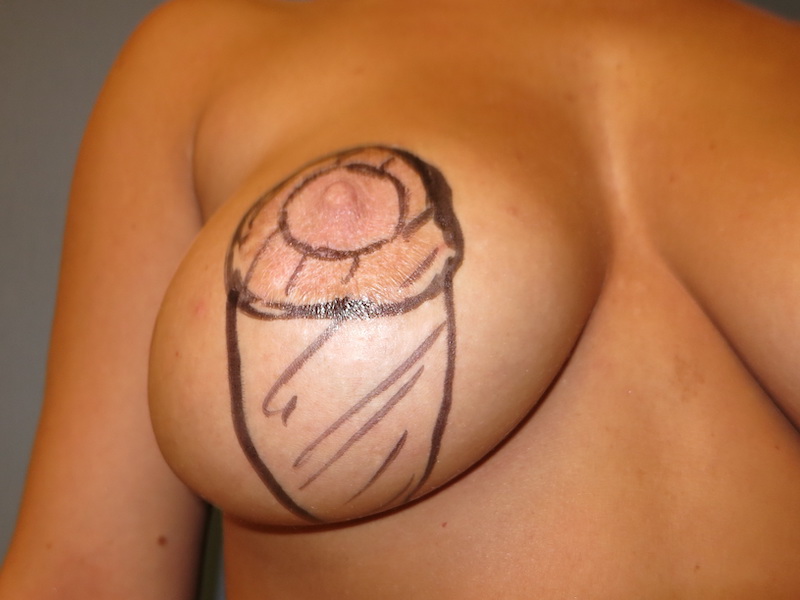
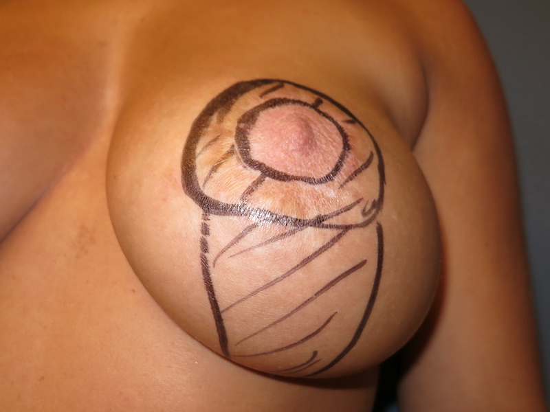
An in-office procedure under local anesthesia was planned. See the last set of images in the before and after series to see the surgical markings and the impressive amount of lower pole breast skin that was removed during surgery. Excess skin was removed and redraped around this patient’s breast tissue.
Follow up images are shown 4 months after surgery. Her areolas are now a more normal size and the bottom-heavy appearance of her breasts are improved. Scars will fade over the next year and her areolas will wake up with time and constrict normally, further improving her areola appearance.
*All photos are actual patient photographs and are for illustrative purposes only. Individual results may vary.