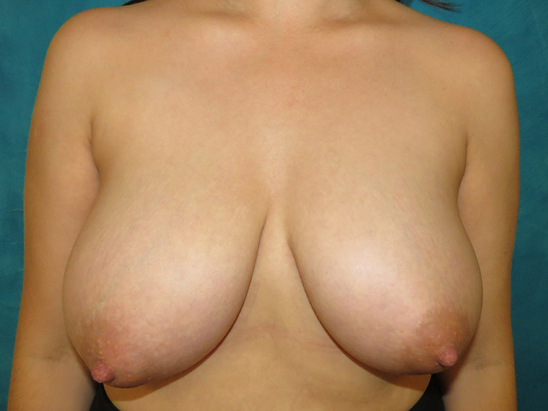
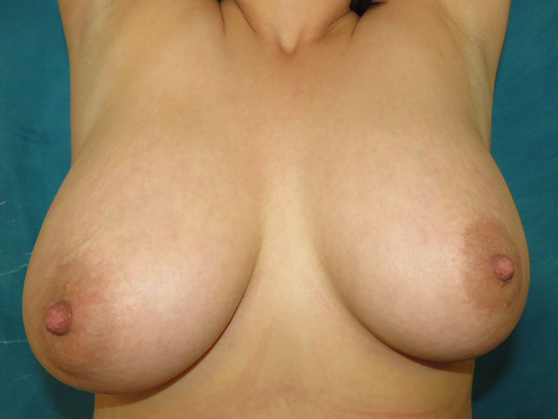
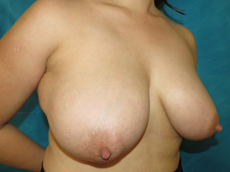
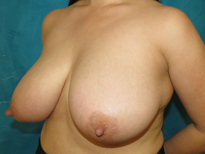
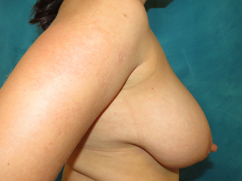
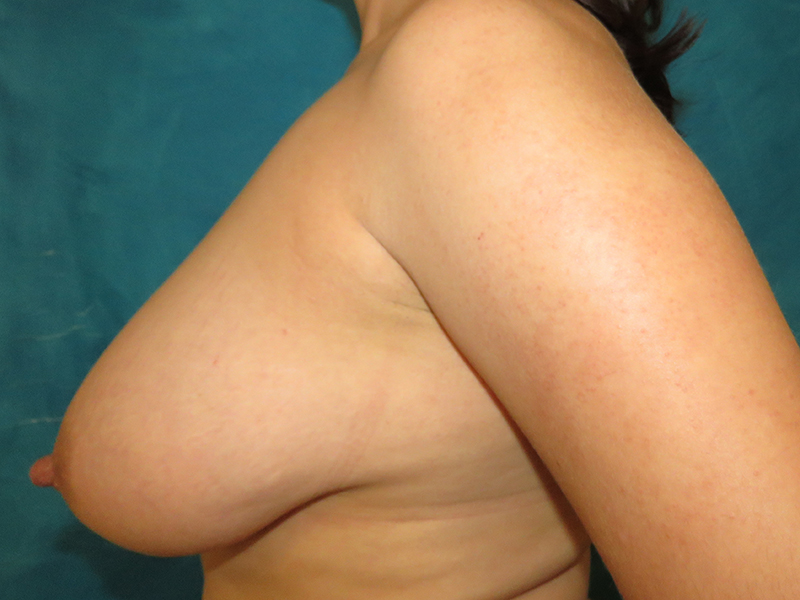
Before and after breast reduction and liposuction of the axillary rolls in a 27 year old woman with “macromastia” and breast asymmetry. She was recently married and was planning on having kids eventually, but wasn’t ready yet. Her goal was to be able to run without pain, wear clothes that didn’t accentuate her chest and to be less top-heavy.
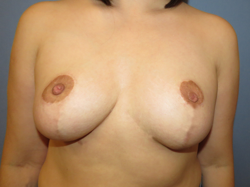
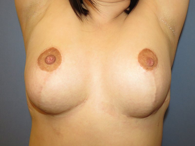
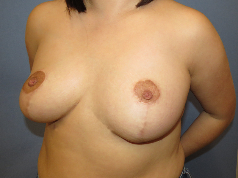
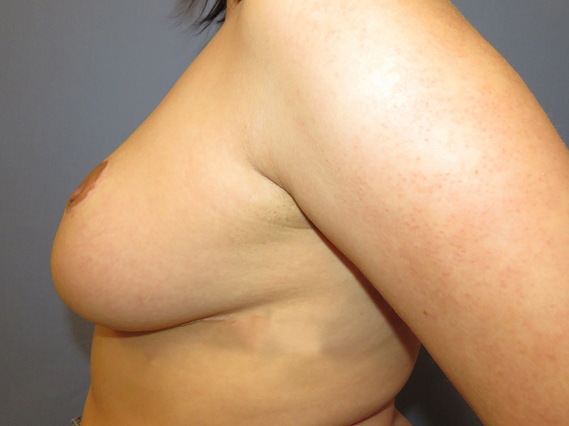
She was elated to learn that our technique of breast reduction does everything possible to preserve the two functions of the breast: breastfeeding ability and erogenous sensation. A robust connection is maintained between the nipple and the remainder of the breast after tissue is removed from the bottom and side of each breast. The nipple and areola is rotated upwards into a pocket created higher up on the chest, creating an “auto-augmentation” effect.
Her breasts extended nearly all the way around to her back, due to excess fat in the bra roll region. In addition to a breast reduction, aggressive liposuction of the bra roll (known as the axillary roll) debulked this area, removing over a liter and a half of fat (think two 750 mL wine bottles filled with fat!).
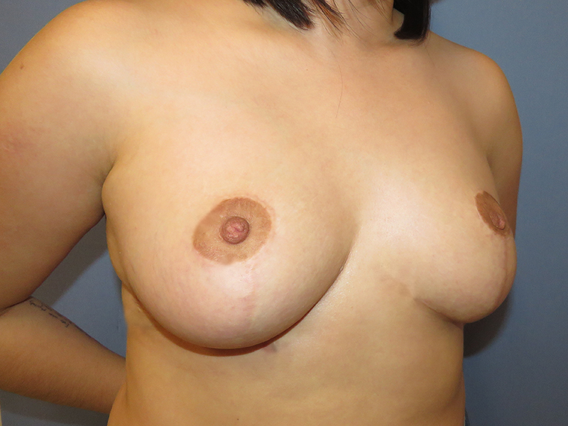
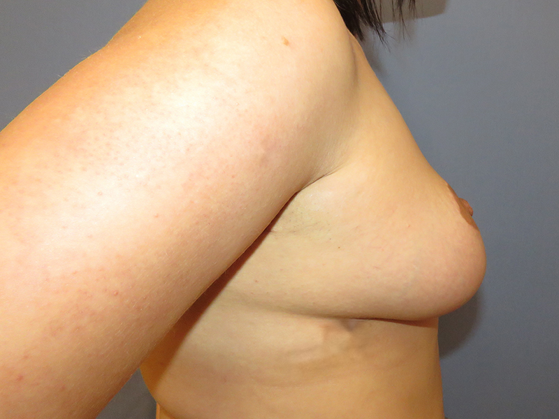
Follow up photos are shown one year after surgery. Her breasts have greatly improved symmetry and she looks much more youthful and proportional with her smaller but proportional breasts. She is very happy with her decision to have surgery and is considering starting her family in the next few years. We have asked her to follow up with us after she has finished having all her children, and to let us know about her breastfeeding success!
*All photos are actual patient photographs and are for illustrative purposes only. Individual results may vary.